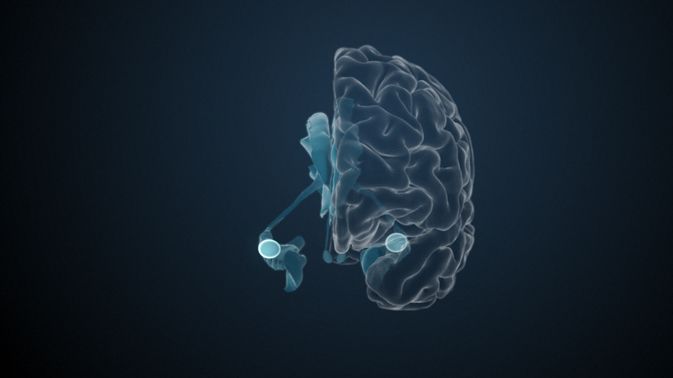
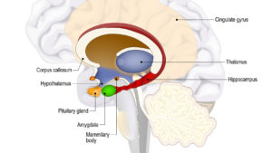
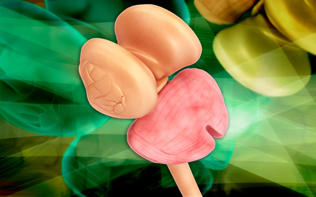
Mastodon
Thalamus

Thalamus

A Central Switch for Everything

The Thalamus is one of those brain areas that crops up in everything – it is considered a central relay station for the brain and therefore is critical to everything we do and think.

thalamus brain neuroleadership


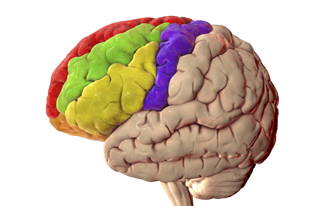
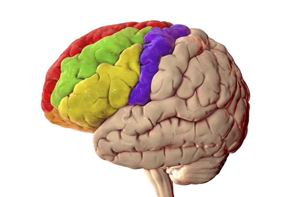
The thalamus is quite unusual in that it is a large brain area at least in surface because it surrounds one of the ventricles in the brain. You often hear about ventricles in passing, and they would, and will, be worth a review at another time. The ventricles are cavities in the brain filled with brain, or spinal fluid, and essential therefore to brain function – but not having a function, such as passing electrical signals, are therefore only studied by neurologists in any detail. Anyhow the thalamus sits at the top of the brain stem and surrounds the third ventricle and sits at a crucial junction.  

It’s first and foremost function seems to be like a junction, an electrical relay station connecting the brains sensory and motor signals to the brain and body. The thalamus is therefore a highly connected brain region and has direct connections to sensory regions, excepting the olfactory region (and interesting observation and may be why the sense of smell is, actually, and amazingly, the fastest sense of all.

brainstem thalamus brain neuroleadership


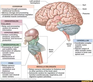
But it doesn’t just do sensory and motor control it also connects to associative parts of the brain and limbic centres so in effect function as a central station for majority of cognitive functions. These are:

  1. Reticular and intralaminar nuclei dealing with arousal and pain regulation
  2. Sensory nuclei regulating all sensory domains except olfaction
  3. Effector nuclei governing motor language function
  4. Associative nuclei connoting cognitive functions
  5. Limbic nuclei encompassing mood and motivation

Given that the thalamus is involved in so much it is almost strange that it does not get more press. The amygdalae have become superstars because of their role in fear and emotion processing.

Even more so when we consider that, as we mentioned above, the thalamus is involved in pain and arousal, pretty important functions, but also wakefulness and alertness.

In fact, the thalamo-cortico-thalamic circuits are though to be heavily involved in consciousness itself – it must be – after all the integration of sensory input into the cerebrum goes directly through the thalamus. Maybe its function is too diffuse and too non-specific to be a clear centre for anything spectacular – those parts of the brain which have clear functions seem to attract more attention and research. But we do also know that damage to the thalamus gives significant risk of coma.

So, it remains that the thalamus is one of the critical brain areas through which just about everything in the brain passes for processing – so we should probably be a bit more thankful for it than we are!

References

Habas, C., Manto, M., and Cabaraux, P. (2019). The Cerebellar Thalamus. Cerebellum 18. doi:10.1007/s12311-019-01019-3.

Haber, S. N., and Calzavara, R. (2009). The cortico-basal ganglia integrative network: The role of the thalamus. Brain Res. Bull. 78, 69–74.

Hwang, K., Bertolero, M. A., Liu, W. B., and D’Esposito, M. (2017). The human thalamus is an integrative hub for functional brain networks. J. Neurosci. 37. doi:10.1523/JNEUROSCI.0067-17.2017.

Redinbaugh, M. J., Phillips, J. M., Kambi, N. A., Mohanta, S., Andryk, S., Dooley, G. L., et al. (2020). Thalamus Modulates Consciousness via Layer-Specific Control of Cortex. Neuron 106. doi:10.1016/j.neuron.2020.01.005.

Wolff, M., and Vann, S. D. (2019). The cognitive thalamus as a gateway to mental representations. J. Neurosci. 39. doi:10.1523/JNEUROSCI.0479-18.2018.

Yen, C. T., and Lu, P. L. (2013). Thalamus and pain. Acta Anaesthesiol. Taiwanica 51. doi:10.1016/j.aat.2013.06.011.

Hormones

Hormones

Hormones and the Brain

A short primer to understand differences between transmitters and hormones and how hormones are directly controlled through the brain.

hormones brain neuroleadership


Hormones are often used colloquially to refer to the changes teenagers go though in adolescence and in reference to sex hormones, often in combination with teenagers. Yes, and there are large impacts on sexual function and related aspects such as fertility and pregnancy. But this interplay with hormones is strongly regulated by the brain, and for the brain, in a loop with stimulation, and secretion of various hormones.

First off, what is a hormone and how do these differ to neurotransmitters and modulators?

Hormones are chemical messengers, communicating to different parts, and organs, of your body. They travel in your bloodstream to tissues or organs. They work slowly, over time, and affect many different processes, including:

  • Growth and development
  • Metabolism – how your body gets energy from the foods you eat
  • Sexual function
  • Reproduction
  • Mood

Endocrine glands, which are special groups of cells, make hormones. The major endocrine glands are the pituitary, pineal, thymus, thyroid, adrenal glands, and pancreas. In addition, men produce hormones in their testes and women produce them in their ovaries.

So, what is the difference to neurotransmitters? Neurotransmitters are released in neurons at the synaptic junctions. They are the messenger between neurons. The most common by far are glutamate and GABA. However, there are also modulators which affects groups of neurons and often act through secondary messengers. Hormones can also be transmitters and modulators. For example, oxytocin, already outlined in lbR-2021-06 is a hormone, a neuromodulator, and a neurotransmitter. This means its effects can be quick, over longer times, and impact many organs in the body.

From this we can see the importance of hormones on brain function and indeed many of the key sites that trigger hormone release sit in the brain notably the pineal gland, the pituitary gland, and the hypothalamus, sitting next to the previously described thalamus. These create feedback loops with the brain and body. In fact, this is a very good reason not to see the brain and body as completely separate entities but as part of a brain-body system, or more accurately a brain-body-environment system.

And which specific hormones are released or triggered from the brain?

Hypothalamus

  • Kisspeptin
  • Oxytocin
  • Gonadotrophin Releasing Hormone

Pineal

  • Melatonin
  • Serotonin

Pituitary

  • Adrenocorticotropic Hormone (ACTH)
  • Growth Hormone
  • Human Chorionic Gonadotropin
  • Luteinizing Hormone
  • Prolactin

Each of these is fascinating in their own right but what you will notice is that a few are probably well known such as Melatonin involved in sleep wake cycles, or oxytocin as we have reviewed previously. Others are more obscure, and others create loops such as ACTH which is triggered by stress reactions stimulating the adrenal gland on the liver to release cortisol which in turn can also have dramatic consequences on brain function.

For this short review it is important to understand that hormones operate within and without the brain. They trigger circuits in the body which in turn influences the brain, or further circuits in the brain which also influences the body – and each of these can have dramatic impacts on function, and notably on long-term health. And this is particularly important over our lifetimes and no less so in older age.

References

Essential guide to hormones: www.shorturl.at/pyzDJ

Hormones: Communication between the Brain and the Body: www.shorturl.at/xAC59

McEwen, B. S. (2020). Hormones and behavior and the integration of brain-body science. Horm. Behav. 119. doi:10.1016/j.yhbeh.2019.104619.

Norepinephrine

Norepinephrine

Colloquially called adrenaline – a powerful activator in the brain

When we speak of adrenaline we think of high stress situations, positive and often negative. Norepinephrine is the neurotransmitter that is related to adrenaline, but not to be confused with the hormone, and it is related to attention, action, but also plasticity and learning.

adrenaline brain

The Sabre-Toothed Tiger jumps out from behind a bush and turns its large yellow eyes onto our friendly cavemen. The tiger gives a low rumbling growl and takes a cautious small step forward, seemingly ready to pounce at an instant. Our caveman, shocked, stands focusing on the tiger his whole body has been rocketed into a high state of alert and tension. His heartbeat has accelerated, his pupils have dilated, his senses have all pricked up and he has laser sharp vision. At this precise moment he is frozen waiting for the slightest abrupt movement which will spur his body into action. Either to launch is rudimentary spear at the tiger or evade and try to escape.

This is often how the primitive roots of our flight or fight response are portrayed – with a threat scenario. Slightly unrealistically: Neolithic man did not live in the same time period as sabre-toothed tigers, and we are adapted to live in the savannah, more likely, than the jungle. Nevertheless, it is easy to imagine, and we have all experienced these periods of shock or tension from simple activities like being surprised by a person jumping from behind a wall, to having a near car crash, to receiving shocking news. Our system activates and all sorts of bodily functions kick off a string of automated reactions. They sympathetic nerve system preparing the system, in very short periods of time, for heightened vigorous activity.

Adrenaline is usually associated with this, it is. But I’d like to give you a brief review of another major neurotransmitter and modulator, norepinephrine also known as noradrenaline. Whereas adrenaline is the hormone signalling responses across the body stimulated by the hypothalamic-pituitary-adrenocortical system norepinephrine is the neurotransmitter that may or may not be associated with the adrenaline response. Chemically they are similar.

norepinephrine brain

So, what does norepinephrine do in the brain? Well, if we take a quick sidestep into ADHD, we can see that there are a number of approaches to deal with ADHD but there are two main classes of drugs to help with attention deficits. One is related to the dopamine system which includes the famous, or infamous, Ritalin, a selective dopamine reuptake inhibitor. The others are related to stimulation and often target the norepinephrine system such as atomoxetine.

Norepinephrine is released mostly in the brain stem specifically in the Locus Coeruleus. Some of you may remember that this is an area that we focused on way back in lbR-2021-01 and is involved in attention. This norepinephrine circuit basically projects throughout the whole cortex as you can see from the diagram above. However, what you will also notice is that there are some similarities to Dopamine which I reviewed in last month’s issue lbR-20201-09.

So, the question is are dopamine and norepinephrine similar, different, or collaborative in their function?

The answer seems to be an irrevocable – probably interrelated! A review in 2020 noted how their functions are similar and they seem to operate in parallel on similar topics – however, we saw last month that dopamine is very strongly involved in the encoding of reward and motivation. It is therefore suitable to conjecture that adrenaline has an arousal and attention function and this complements dopamine. This thereby also stimulates encoding of significance be that of positive or particularly of negative and stressful events which are particularly powerful in the brain. The review also notes norepinephrine’s importance in plasticity and how this also functions in parallel with dopamine.

So, in summary, norepinephrine is a key transmitter that has wide-reaching impacts on the brain and operates closely with dopamine to guide attention, encode and interpret emotional significance, and in guiding learning

References

Norepinephrine

Mather, M., Clewett, D., Sakaki, M., and Harley, C. W. (2016). Norepinephrine ignites local hotspots of neuronal excitation: How arousal amplifies selectivity in perception and memory. Behav. Brain Sci. 39. doi:10.1017/S0140525X15000667.

Moret, C., and Briley, M. (2011). The importance of norepinephrine in depression. Neuropsychiatr. Dis. Treat. 7. doi:10.2147/NDT.S19619.

Saboory, E., Ghasemi, M., and Mehranfard, N. (2020). Norepinephrine, neurodevelopment and behavior. Neurochem. Int. 135. doi:10.1016/j.neuint.2020.104706.

Schwarz, L. A., and Luo, L. (2015). Organization of the locus coeruleus-norepinephrine system. Curr. Biol. 25. doi:10.1016/j.cub.2015.09.039.

van der Linden, D., Tops, M., and Bakker, A. B. (2021). The Neuroscience of the Flow State: Involvement of the Locus Coeruleus Norepinephrine System. Front. Psychol. 12. doi:10.3389/fpsyg.2021.645498.

Dopamine vs Norepinephrine

Ranjbar-Slamloo, Y., and Fazlali, Z. (2020). Dopamine and Noradrenaline in the Brain; Overlapping or Dissociate Functions? Front. Mol. Neurosci. 12. doi:10.3389/fnmol.2019.00334.

The Amygdala

The Amygdala

Fear, or emotions, or attention?

The Amygdala is one of those brain areas that gets a lot of attention. A lot. In fact, it may be one of the most famous areas of the brain – in no short part due to its role in fear and what Daniel Goleman called the “Amygdala Hijack” to describe situations in which emotionality takes over your brain – or supposedly at least. But the amygdala is a little misrepresented – let’s clear up its reputation.

amygdala brain

SM are the initials of a patient, who according to the case in a well-cited paper by Feinstein et al. in 2010, exhibits little to no fear. What is special about SM is that she has severe damage to both her Amygdalae. And though at the time there was known to be a strong relationship between fear and Amygdala function, SM’s rare condition enables the study of this in real world scenarios.

She was put through a series of situations and her fear measured. For example, she had consistently expressed a fear of spiders and snakes in previous interviews and so she was taken to an exotic pet store and presented with snakes and spiders to document her response. Surprisingly despite her insistence she is “afraid “ of these, she showed no fear at all. In fact, she showed the opposite: curiosity, reaching out to touch the snakes and spiders and stroke them!

In addition to this she was taken to, ostensibly, America’s most haunted house. The Waverly Hills Sanatorium Haunted House, showing no fear on a tour in contrast to other participants, and researchers, on the same tour. Her fear and emotional response were also collected systematically in everyday life with an emotional diary and through structured interviews. She indeed seemed to show no fear or to experience threat.

One notable experience also points to a key role of the Amygdala or absence of this in SM. She lived in a city and one of her walking routes home passed through a parking lot. On one occasion she was held up at knifepoint and robbed. For many this may have been a traumatic experience – but moreover it would almost certainly have led to avoiding this parking lot or walking home at night through this place. Also, the place would be expected to trigger negative memories. Not so with SM. She continued to walk across the parking lot unabated and unaffected by her negative experience there. This shows a key role of the Amygdala, and maybe underestimated with the focus on fear processing, namely that of learning.

In fact, a recent study did just this. They attempted to connect the amygdala to learning and not through triggering the emotional impact. Emotions trigger large networks in the brain and so it is difficult to disentangle the effects of different regions in the brain. In these experiments by Bass et al. in 2021 they used Deep Brain Stimulation to simulate the Amygdala (or not) in rats when encountering objects. They showed that the stimulation increased the memory of objects without observably activating emotions.

fear brain amygdala

Illustration such as this are used in research into Fear – just looking at this picture consistently shows activation patterns in the amygdala.

Illustration such as this are used in research into Fear – just looking at this picture consistently shows activation patterns in the amygdala.[/caption]This puts the Amygdala at the crossroads of memory and learning and particularly Pavlovian conditioning – so relating positive or aversive stimuli with contexts. This was clearly missing in SM whose experience in the parking lot did not lead to her having aversive reactions to said parking lot. Though we have outlined this clear relationship to fear there have been just as many relationships to positive emotions and the Amygdala. This is why many researchers see it as a salience processing unit rather than a fear centre. It shows us what is important and hence where to focus our attention, or not, and what to learn or not.

However, though there is also strong activation in the Amygdala to positive or appetitive cues, cases such as in SM do not show much dysfunction of positive emotions. So, it seems the Amygdala is involved in all emotional processing, but rewarding and positive experiences rely on other networks whereas fear and threat are strongly related to, or even dependent on, the Amygdala.

This key function of the Amygdala can be seen in its location and connections: it sits in the limbic system close to the hippocampus, itself considered a memory centre, but also close to the thalamus and prefrontal cortex. It is split into different subsections, simply three. The medial, the middle bit, is connected strongly to the olfactory centres (smell), the basolateral, to the frontal and cerebral cortex, and the central & anterior to the brain stem, hypothalamus, and sensory centres.

brain amygdala

Amygdala and neighbouring structures


All of these seem to make sense – connecting sensory input to emotional and hormone release in the hypothalamus, and to the frontal regions to control attention and close to the hippocampus to guide learning, not to mention sensory centres to build associations. Of notable interest, and little discussed is the connections to the olfactory centre. Though the majority of research is into visual stimuli, particularly of faces to which the amygdala can be more responsive than other areas of the brain supposedly specialised in faces (the FFA reviewed in lbR-2021-08).

A recent piece of research shows why – the sense of smell is one of our fastest systems to respond – harking back to a time when our sense of smell was much more important in everyday life and more than in modern society. Iravani et al. from the Karolinska Institute in Sweden showed in a recent piece of research that the olfactory system is a high-speed circuit and particularly to aversive smells which influence our avoidance behaviours. This is often also unconscious.

So, this paints a clearer picture of the function of the Amygdala as an emotional attention centre that is especially reactive and necessary for threat and aversive contexts and drives learning, memory, and conditioning. However, the role of the Amygdala may have unintentionally been tainted by Daniel Goleman’s 1995 description of the “Amygdala Hijack”. This term has been used by many an aspiring neuroleadership expert or coach.

Simply put, Goleman, at the time, painted picture whereby the amygdala reaction essentially hijacked the rational brain and rendered us at the whims of our emotionality. This is an oversimplified description which is appealing to a broader audience but does have some truth in it. Even as an aspiring neuroleadership expert I found the term a little oversimplified – however, it was also useful to describe to lay audiences how emotionality can take over the brain. The fact is, threat is a basic survival instinct and so can, and will, activate many stress systems in the body. However, how to engage and deal with this and is not clearly identified with the term amygdala hijack.

The SCOAP model that regular readers will be familiar with, goes some way to explain this in more detail – Firstly, different concepts can trigger a negative reaction (e.g. self-esteem threat but also loss of control or loss of orientation). Secondly, this can be very individualised. Thirdly, some people respond to threats much stronger than others. Fourthly, many of these are also conditioned responses. Fourthly we are looking at broad networks in the brain and body. So yes, the Amygdala hijack could be a way to describe the emotional response we have, but is too simplified to be useful and misconstrues how the amygdala and brain functions together. SCOAP is a much better way to formulate this and explore – for those who want to learn more.

But back to our two almond-shaped structures in our brain. They are widely researched, seem to have an oversized influence on brain functions, do activate strongly to threat and fear, and control response to this, and control aversive and avoidance behaviours – in fact so much so that political affiliation can be accurately predicted by looking at amygdala activation as I outline in the earlier article on fear in society.

So, the amygdala it is a powerful brain region related to primal networks – and also positive learning, and sometimes being afraid and cautious of threat is very good thing. As SM didn’t learn, sometimes it is good to avoid dangerous places. But too much fear is not good thing either. So, we may need to use our prefrontal to exert some top-down influence on our amygdala or at least refocus on positives. And that will also lower stress. And lead to higher wellbeing

References

Case of SM

Feinstein, J. S., Adolphs, R., Damasio, A., and Tranel, D. (2010). The Human Amygdala and the Induction and Experience of Fear. Curr. Biol. 21, 1–5. doi:10.1016/j.cub.2010.11.042.

Olfactory response

Iravani, B., Schaefer, M., Wilson, D. A., Arshamian, A., and Lundström, J. N. (2021). The human olfactory bulb processes odor valence representation and cues motor avoidance behavior. Proc. Natl. Acad. Sci. U. S. A. 118. doi:10.1073/pnas.2101209118.

Amygdala and memory and learning

Bass, D. I., Partain, K. N., and Manns, J. R. (2012). Event-specific enhancement of memory via brief electrical stimulation to the basolateral complex of the amygdala in rats. Behav. Neurosci. 126. doi:10.1037/a0026462.

Article on DANA website: https://www.dana.org/article/beyond-emotion-understanding-the-amygdalas-role-in-memory/

Steinberg, E. E., Gore, F., Heifets, B. D., Taylor, M. D., Norville, Z. C., Beier, K. T., et al. (2020). Amygdala-Midbrain Connections Modulate Appetitive and Aversive Learning. Neuron 106. doi:10.1016/j.neuron.2020.03.016.

Review

LeDoux, J. (2007). The amygdala. Curr. Biol. 17, R868-74. doi:10.1016/j.cub.2007.08.005.

Amygdala and fear

Hardee, J. E., Thompson, J. C., and Puce, A. (2008). The left amygdala knows fear: laterality in the amygdala response to fearful eyes. Soc. Cogn. Affect. Neurosci. 3, 47–54.

Murray, E. A. (2007). The amygdala, reward and emotion. Trends Cogn. Sci. 11, 489–497. doi:10.1016/j.tics.2007.08.013.

Michely, J., Rigoli, F., Rutledge, R. B., Hauser, T. U., and Dolan, R. J. (2020). Distinct Processing of Aversive Experience in Amygdala Subregions. Biol. Psychiatry Cogn. Neurosci. Neuroimaging 5. doi:10.1016/j.bpsc.2019.07.008.

Behaviour and stress

Zhang, W. H., Zhang, J. Y., Holmes, A., and Pan, B. X. (2021). Amygdala Circuit Substrates for Stress Adaptation and Adversity. Biol. Psychiatry 89. doi:10.1016/j.biopsych.2020.12.026.

Yang, Y., and Wang, J. Z. (2017). From structure to behavior in basolateral amygdala-hippocampus circuits. Front. Neural Circuits 11. doi:10.3389/fncir.2017.00086.

Gründemann, J., Bitterman, Y., Lu, T., Krabbe, S., Grewe, B. F., Schnitzer, M. J., et al. (2019). Amygdala ensembles encode behavioral states. Science (80-. ). 364. doi:10.1126/science.aav8736.

Krabbe, S., Gründemann, J., and Lüthi, A. (2018). Amygdala Inhibitory Circuits Regulate Associative Fear Conditioning. Biol. Psychiatry 83. doi:10.1016/j.biopsych.2017.10.006.

Šimić, G., Tkalčić, M., Vukić, V., Mulc, D., Španić, E., Šagud, M., et al. (2021). Understanding emotions: Origins and roles of the amygdala. Biomolecules 11. doi:10.3390/biom11060823.

Kim, J., Zhang, X., Muralidhar, S., LeBlanc, S. A., and Tonegawa, S. (2017). Basolateral to Central Amygdala Neural Circuits for Appetitive Behaviors. Neuron 93. doi:10.1016/j.neuron.2017.02.034.

The Prefrontal Cortex

The Prefrontal Cortex

The part of the brain that is considered particularly human

The frontal cortex gets a lot of press being known as the executive centre and though it is a large and complex area, I believe a short review is necessary to clarify what it does do.

brain prefrontal

The frontal or prefrontal cortex gets a lot of publicity, and quite rightly so. It is considered our “executive centre”, the part of the brain that does all that high-level stuff that makes us very human. It is also the part of the brain that is vastly different to chimpanzees and one that we can see has developed over the human lineage.

I have already reviewed certain parts that are, or can be considered, a part of the prefrontal cortex in previous issues, namely the orbitofrontal cortex in lbR-2021-03 and also the anterior cingulate cortex in lbR-2021-04, but also in lbR-2021-06 when I looked at the social brain. But this issue I would like to take a step back and give you a brief overview of the prefrontal which will enable us to look at different regions in more detail in the future.

When we talk of the prefrontal we are, obviously, talking about the frontal lobe of the brain. However, this can be defined in different ways:

They cytoarchitectonic approach defines the prefrontal as the frontal part of the brain that has a granular layer IV. Which may sound like mumbo-jumbo to the non-specialist. The cortex is structured into layers of neurons normally having six layers (I reviewed in lbR-2021-01). However, this is strictly not true with some traditionally classed in the prefrontal not having a granular layer IV.

The projection zone defines the prefrontal cortex as the area that is connected, or has projections from, the mediodorsal nucleus of the thalamus. Though widely accepted more recent and advanced imaging has shown that this is also strictly not true.

The electrically silent area of the prefrontal cortex is another way to define the prefrontal as the area in front of the motor cortex that does not stimulate and observable movements when electrically stimulated. This helps distinguish prefrontal areas from motor areas but electrically silent is the opposite of what is happening in the prefrontal with wide-reaching brain activation on stimulation of the prefrontal (showing its importance).

The above are of passing interest only to us as non-neuroscientists. Of more interest are the specific regions. These are first split into the medial (inside), lateral (outside), and orbital (underside) surfaces.

prefrontal brain

From Holt et al. 2008

The medial surfaces are those that are “in the fold” between the hemispheres. This is often generally referred to as the Medial Prefrontal Cortex (32 above), Ventromedial Prefrontal Cortex (25 above), and the Anterior Cingulate Cortex (24 above) is normally classed separately though sometimes included as part of the medial prefrontal cortex. All of these can be split into smaller regions.

The lateral surfaces are normally split into three larger regions. The Dorsolateral Prefrontal Cortex (9,10, 46 above), the Ventrolateral Prefrontal Cortex (44, 45, 47 above), and the Orbitofrontal Cortex which is actually the bottom of the Prefrontal Cortex.

So, what does the prefrontal cortex do?

“The PFC is a large and complex brain region that is best conceptualized as being located at the highest point of a sensorimotor pyramid, starting with the primary sensory cortices and ending at the primary motor cortex (Figure 71-3). Highly processed information from sensory association areas converges onto the PFC, which then integrates the information with existing priorities, leading to the construction of adaptive behavioral plans based on this input.”

– Holt et al. 2008

We often talk about executive functions, but that term also needs clarifying.

  1. Top-down processes whereby we are exerting control on bottom-up processes, so, for example, exercising restraint.
    • Inhibition
    • Conscious cognitive effort such as doing calculations
    • Overcoming desire to do something else, e.g., moving when you don’t feel like moving
  1. Attention and memory
    • Focused attention or attending to stimuli – consciously or automatically
    • Memory especially short-term or working memory i.e. keeping something in mind so that you can work with it immediately
  1. Social processing
    • Empathy and social models associated with medial prefrontal cortex as reported in lbR-2021-06
  1. Decision making
    • Decision making including analysis
    • Value based judgements balancing emotion and reason especially in the orbitofrontal cortex as reported in lbR-2021-03
    1. Planning and Strategy
    • Planning and strategy – or adaptive behavioural plans based in input and value-based projections
  1. Speech production and language
    • Various aspects of language processing are related to prefrontal areas related to speech production, language comprehension, and response planning before speaking which include and are related to the above functions such as mediated implicit memory retrieval of verbs or noun recollection. These functions differ in the hemispheres.

However, despite the above pointing to the consolidatory and high-order, and top-down functions it must also be noted that some “lower-order” functions such as sleeping with degradation in the prefrontal associated with lowered sleep quality. So as usual we can see that it is hard to clearly delineate the brain as I continually stress it is an integrated whole.

Another important aspect of the prefrontal is that of the role of dopamine that a review in the next article. As you will see dopamine can be considered an attention and motivational molecule and it is therefore no surprise that the prefrontal is rich in dopamine receptors and the meso-cortic pathway is a major dopamine highway.

More recent research has also shown that other brain areas such as the cerebellum also contributes to cognitive processes – but this is likely in the form of immediate feedforward processes based on building cognitive maps – so specialised habits rather than effortful thought and top-down processing.

So, we can see that the prefrontal cortex is indeed involved in a lot of things that make us very human and differentiate us from other animals. It is not the only region that contributes to this but its certainly seems to be a consolidated point of the brain. Without which I would certainly not have been able to exert the control, planning, inhibition, and attention needed to write this article. And you wouldn’t have been able to keep your attention on this article to get this far

References

Bang, D., and Fleming, S. M. (2018). Distinct encoding of decision confidence in human medial prefrontal cortex. Proc. Natl. Acad. Sci. U. S. A. 115. doi:10.1073/pnas.1800795115.

Bechara, A. (2000). Characterization of the decision-making deficit of patients with ventromedial prefrontal cortex lesions. Brain 123, 2189–2202. doi:10.1093/brain/123.11.2189.

Bicks, L. K., Koike, H., Akbarian, S., and Morishita, H. (2015). Prefrontal cortex and social cognition in mouse and man. Front. Psychol. 6. doi:10.3389/fpsyg.2015.01805.

Carlén, M. (2017). What constitutes the prefrontal cortex? Science (80-. ). 358. doi:10.1126/science.aan8868.

Dolan, R. J. (2007). The human amygdala and orbital prefrontal cortex in behavioural regulation. Philos. Trans. R. Soc. London – Ser. B Biol. Sci. 362, 787–99. doi:10.1098/rstb.2007.2088.

Donahue, C. J., Glasser, M. F., Preuss, T. M., Rilling, J. K., and Van Essen, D. C. (2018). Quantitative assessment of prefrontal cortex in humans relative to nonhuman primates. Proc. Natl. Acad. Sci. U. S. A. 115. doi:10.1073/pnas.1721653115.

Duverne, S., and Koechlin, E. (2017). Rewards and Cognitive Control in the Human Prefrontal Cortex. Cereb. Cortex 27. doi:10.1093/cercor/bhx210.

Dux, P. E., Tombu, M. N., Harrison, S., Rogers, B. P., Tong, F., and Marois, R. (2009). Training improves multitasking performance by increasing the speed of information processing in human prefrontal cortex. Neuron 63, 127–138.

Holt, D. J., Öngür, D., Wright, C. I., Dickerson, B. C., and Rauch, S. L. (2008). “Neuroanatomical Systems Relevant to Neuropsychiatric Disorders,” in Massachusetts General Hospital Comprehensive Clinical Psychiatry doi:10.1016/b978-0-323-04743-2.50073-1.

Jenkins, A. C., and Mitchell, J. P. (2011). Medial prefrontal cortex subserves diverse forms of self-reflection. Soc. Neurosci. 6, 211–8.

Jeon, H.-A., and Friederici, A. D. (2013). Two principles of organization in the prefrontal cortex are cognitive hierarchy and degree of automaticity. Nat. Commun. 4, 2041. doi:10.1038/ncomms3041.

Kim, H., Shimojo, S., and O’Doherty, J. P. (2011). Overlapping responses for the expectation of juice and money rewards in human ventromedial prefrontal cortex. Cereb. Cortex 21, 769–776.

Lara, A. H., and Wallis, J. D. (2015). The role of prefrontal cortex in working memory: A mini review. Front. Syst. Neurosci. 9. doi:10.3389/fnsys.2015.00173.

Likhtik, E., Pelletier, J. G., Paz, R., and Pare, D. (2005). Prefrontal Control of the Amygdala. 25, 7429–7437. doi:10.1523/JNEUROSCI.2314-05.2005.

Massi, B., Donahue, C. H., and Lee, D. (2018). Volatility Facilitates Value Updating in the Prefrontal Cortex. Neuron 99. doi:10.1016/j.neuron.2018.06.033.

Michel, M., and Morales, J. (2020). Minority reports: Consciousness and the prefrontal cortex. Mind Lang. 35. doi:10.1111/mila.12264.

Ott, T., and Nieder, A. (2019). Dopamine and Cognitive Control in Prefrontal Cortex. Trends Cogn. Sci. 23. doi:10.1016/j.tics.2018.12.006.

Perry, A., Saunders, S. N., Stiso, J., Dewar, C., Lubell, J., Meling, T. R., et al. (2017). Effects of prefrontal cortex damage on emotion understanding: EEG and behavioural evidence. Brain 140. doi:10.1093/brain/awx031.

Petrides, M. (2005). Lateral prefrontal cortex: Architectonic and functional organization. Philos. Trans. R. Soc. B Biol. Sci. 360. doi:10.1098/rstb.2005.1631.

Raine, a, Lencz, T., Bihrle, S., LaCasse, L., and Colletti, P. (2000). Reduced prefrontal gray matter volume and reduced autonomic activity in antisocial personality disorder. Arch. Gen. Psychiatry 57, 119–27; discussion 128-9.

Smaers, J. B., Gómez-Robles, A., Parks, A. N., and Sherwood, C. C. (2017). Exceptional Evolutionary Expansion of Prefrontal Cortex in Great Apes and Humans. Curr. Biol. 27. doi:10.1016/j.cub.2017.01.020.

Starkweather, C. K., Gershman, S. J., and Uchida, N. (2018). The Medial Prefrontal Cortex Shapes Dopamine Reward Prediction Errors under State Uncertainty. Neuron 98. doi:10.1016/j.neuron.2018.03.036.

Wang, J., Kurth-Nelson, Z., Kumaran, D., Tirumala, D., Soyer, H., Leibo, J., et al. (2018). Prefrontal Cortex as a Meta-Reinforcement Learning System. Prefrontal cortex as a meta-reinforcement Learn. Syst. doi:10.1101/295964.

Xu, H., Liu, L., Tian, Y., Wang, J., Li, J., Zheng, J., et al. (2019). A Disinhibitory Microcircuit Mediates Conditioned Social Fear in the Prefrontal Cortex. Neuron 102. doi:10.1016/j.neuron.2019.02.026.

Manly Testosterone

Manly Testosterone

. . .


This content is only for Premium Brains - please subscribe to access this. All subscriptions come with a free trial.
Register for free to receive access to "Healthy Brains" and other selected articles.
Subscribe to Premium Brain (monthly or annual) to access all articles and access the download page.

Subscribe now


leadership brain magazine